
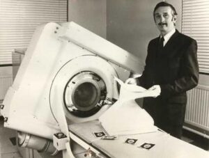
In the mid-1960s, British engineer Godfrey Hounsfield conceived a bold and visionary idea: could we “look inside a box without opening it”? His inspiration came from pondering how cosmic rays might reveal hidden chambers within Egypt’s pyramids. But instead of exploring ancient tombs, Hounsfield turned his attention to the human skull—a “box” that protects the brain.
Recognizing that conventional X-ray imaging could not clearly depict the soft tissues of the brain, Hounsfield proposed a groundbreaking approach: slicing the brain into virtual layers, scanning each one with focused X-ray beams, and using a computer to reconstruct the internal density point by point. This became the foundation of Computed Tomography (CT scanning).
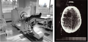
With little funding, Hounsfield built his first prototype using scrap materials. Thanks to the quiet support of a forward-thinking executive at EMI, he installed the first CT scanner at Atkinson Morley Hospital in London. On October 1, 1971, the first CT image of the human brain—revealing a tumor in the left frontal lobe—was produced, marking a turning point in medical history.
Although EMI did not pursue the medical market for long, Hounsfield’s invention paved the way for major players like GE and Siemens to develop faster, full-body CT scanners with higher resolution. In 1981, Hounsfield was knighted by Queen Elizabeth II and continued his research until the end of his life.
From a dream of “seeing through the skull,” CT scanners have become an essential diagnostic tool—especially in emergency medicine, oncology, and trauma care. By 2020, more than 80 million CT scans were performed annually in the United States alone. It all began with one man’s vision—to reveal the invisible.